Hepa1-6-iRFP-Puro
| Species | Mouse |
| Cell Type | C57/L hepatoma |
| Transgene | Near-infrared fluorescent protein (iRFP; ex/em 690/713nm) |
| Selection Gene | Puromycin resistance (Puro) |
-
Description
Hepa1-6-iRFP-Puro is a polyclonal population of the hepatoma Hepa 1-6 cell line (ATCC® CRL-1830™). To achieve stable reporter expression in the polyclonal population, parental Hepa1-6 cells were transduced with LV-iRFP-P2A-Puro (LV032) and selected using puromycin. LV-iRFP-P2A-Puro encodes the near-infrared fluorescent protein (iRFP) cDNA linked to the puromycin resistance gene (Puro) via a P2A cleavage site (P2A) under the spleen focus-forming virus (SFFV) promoter.
*The ATCC trademark and trade name and any and all ATCC catalog numbers are trademarks of the American Type Culture Collection.
This cell line has been tested for mycoplasma contamination and is certified mycoplasma free.
The parental Hepa1-6 cell line has been authenticated and certified free of interspecies cross contamination by short tandem repeat (STR) profiling with 9 STR loci.
Due to the immunogenicity of the reporter genes in this cell line, we recommend using immunocompromised mice for in vivo studies.
Replication Competent Lentivirus (RCL) Test (including a test report) is available for this cell line at an added cost. Contact us to learn more.
-
Characterization
Morphology
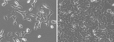 Low and high density cell morphology (200x)
iRFP Expression
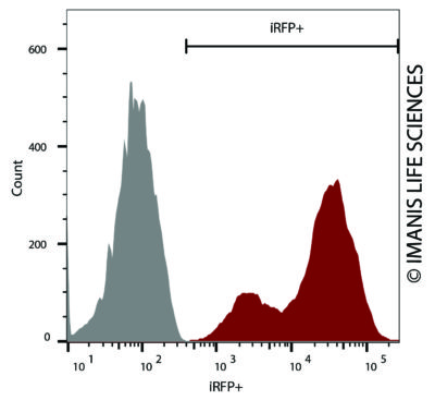 Hepa1-6-iRFP-Puro (red) or control (Hepa1-6; grey) cells were fixed with paraformaldehyde and analyzed by flow cytometry.
 -
Growth Conditions
Complete Growth Medium: DMEM supplemented with 10% FBS, 1X Penicillin/Streptomycin, and 2 µg/mL puromycin.
The addition of puromycin to the complete growth medium maintains high reporter expression over continued passage of the cells. It is highly recommended, especially if the cells undergo multiple passages prior to being used for studies.
These cells should be grown in the indicated medium and passaged when they reach confluency. For routine passaging, cells are recommended to be split at a 1:10 ratio every 3-4 days.
-
Usage Information
These cells are suitable for in vitro and in vivo experimentation. Hepa1-6 cells form primary tumors and spontaneous lung metastases post implantation into syngenic C57L/J mice1,2. However, tumor formation in syngenic mice is highly variable, so for the most consistent results immunocompromised mice are recommended for studies.
The iRFP transgene facilitates in vivo and ex vivo fluorescence imaging of implanted cells. To reduce background autofluorescence, mice should be fed an alfalfa-free diet for at least a week prior to imaging.
The cells can be amplified in vitro and used to generate additional frozen stocks. Cryopreservation of low passage stocks is recommended. Frozen stocks should be preserved in a designated cryopreservation medium.
These cells were generated via lentiviral vector transduction. The lentiviral vector used for transduction was a self-inactivating (SIN) vector in which the viral enhancer and promoter have been deleted. Transcription inactivation of the LTR in the SIN provirus increases biosafety by preventing mobilization by replication competent viruses and enables regulated expression of the genes from the internal promoters without cis-acting effects of the LTR3. Nevertheless, all work with these cells should be performed under biosafety-level 2 (BSL2) conditions by trained personnel. Institutional requirements may permit handling of these cells under BSL1 conditions if certain criteria are met.
References:
1Lei and Ling. World J Gastroenterol 2015. 21: 10137-10149.
2Wang et al. Cell Physiol Biochem 2016. 38: 306-318.
3Miyoshi et al. J Virol 1998. 72:8150-8157.