
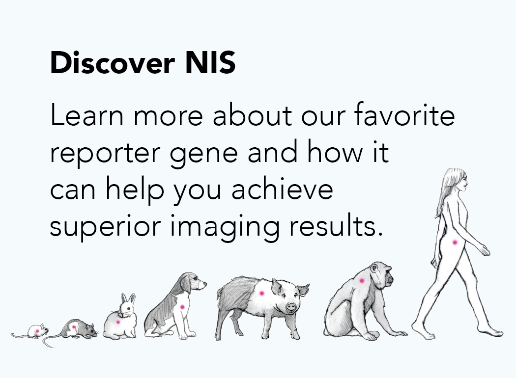
Nuclear Reporters
For noninvasive in vivo nuclear (PET/SPECT) imaging
Description
Nuclear reporters are proteins that trap various radiotracers in the cells in which they are expressed. Most nuclear reporters, including the sodium iodide symporter (NIS), human norepinephrine transporter (hNET), human somatostatin receptor 2 (hSSTR2), and human dopamine receptor D2 (hDRD2), are transporters or receptors that directly mediate uptake of specific radiotracers into the cells. These reporter proteins are endogenously expressed in various human tissues, where they have a diverse range of important functions. Another nuclear reporter, Herpes Simplex Virus Thymidine Kinase (HSVTK), is an enzyme that catalyzes the phosphorylation of intracellular radiolabeled pro-drugs so that they are no longer membrane permeable and are trapped in the cell. For imaging studies, target cells and viruses can be engineered to express high levels of any of the recombinant nuclear reporter proteins.
Because they concentrate radiotracers, nuclear reporters can be used to noninvasively track cells in vivo through nuclear imaging. Imaging with nuclear reporters offers several key advantages: it produces high-resolution 3D images that are fully quantitative, it is translational from small to large animals as well as humans (depending on the reporter), and it can be used for longitudinal imaging studies as species-specific versions exist for some of the reporters.
How to use nuclear reporters for imaging
Reagents: Nuclear reporters trap a variety of different radiotracers.
| Reporter | SPECT radiotracers | PET radiotracers |
|---|---|---|
| NIS | Iodine-123
Iodine-125 [99mTc]-pertechnetate |
Iodine-124 [18F]-TFB |
| hNET | [123I]-MIBG [125I]-MIBG |
[124I]-MIBG [11C]-ephedrine [11C]-mHED |
| hSSTR2 | [111In]-DTPA-octreotide [111In]-pentetreotide |
[68Ga]-DOTA-TATE [18F]-FP-Gluc-TOCA |
| HSVTK | [123I]-FIAU [125I]-FIAU |
[124I]-FIAU [11C]-FIAU [18F]-FIAU [18F]-FHBG |
| hDRD2 | [123I]-Ioflupane |
[18F]-FESP [18F]-Fallypride |
Equipment: SPECT or PET machines can be used to detect radiotracer signal and biodistribution of nuclear reporter-expressing cells or viruses. Combining SPECT or PET with computed tomography (CT) or magnetic resonance (MR) imaging gives the most informative results, as images can be co-registered to give anatomical localization of the radiotracer signal.
Additional considerations: Most of the nuclear reporters are endogenously expressed in certain organs or tissues. When selecting a nuclear reporter, the imaging target site should be considered. Additionally, not all radiotracers may effectively reach certain target tissues/organs. See the chart under the “When to use nuclear reporters” section, below, for more information.
Workflow:
![]()
When to use nuclear reporters
Nuclear reporters are well suited for longitudinal, noninvasive imaging studies in regenerative medicine, gene therapy, oncology, and oncolytic virotherapy, where they can be used to track the biodistribution of cell/virus therapies or the destruction of tumors. Nuclear reporters can be used for small or large animal imaging and most are translatable to the clinic. The following chart gives general guidelines for organs/tissues that are suitable for imaging with the different nuclear reporters:
| Reporter | Endogenous expression | Recommended for | Not recommended for |
|---|---|---|---|
| NIS | Thyroid, stomach, mammary glands, salivary glands | Heart, lungs, liver, kidneys, orthopedics (joints, bone), immune cells | Brain, CNS |
| hNET | CNS and PNS | Brain, CNS, immune cells | Liver, kidneys, intestines |
| hSSTR2 | Cerebrum, kidney | Tracking oncolytic viruses | Kidneys, pancreas, pituitary gland |
| HSVTK | None | Heart, muscle, immune cells | Liver, intestines |
| hDRD2 | Striatal-Nigral System of the brain | Brain and CNS | Liver, kidneys, intestines |